Distribution of phosphorylated α-synuclein in Octodon degus brains.
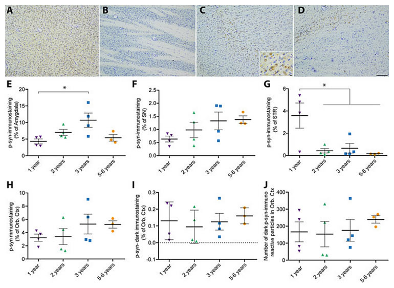
(A–D) Representative images of phosphorylated α-synuclein (p-syn) in different brain regions of 3 years-old octodons (A) Amygdala; (B) Orbito-frontal cortex (Orb Ctx); (C) Striatum (STR); (D) substantia nigra (SN). Inset is a high magnification picture in striatum. (E–J) Bottom panels display surface quantification of the p-syn immunostaining in amygdala (E), SN (F), STR (G), Orb Ctx (H–J). Significant age effect was observed in amygdala [F(3,11) = 4.275, p = 0.0314] and STR [F(3,11) = 5.977, p = 0.0114]. (J) Number of dark spot of p-syn immunostaining in the orbital cortex. Scale bar: 100 μm, high magnification: 10 μm. *p
MORE +